
Start here
Understanding the diagnosis—and the reason for surgery
- A mediastinal mass is an abnormal finding in the central part of the chest. Many are not cancer.
- Thymoma is a tumor of the thymus gland. Treatment depends on its extent and whether it can be completely removed.
- Myasthenia gravis is an autoimmune condition that causes muscle weakness. Removing the thymus can help selected patients, even without a tumor.
The basics
What is the mediastinum?
The mediastinum is the central space in your chest between the lungs. It contains the heart, major blood vessels, windpipe, esophagus, lymph nodes, and thymus gland. The thymus sits behind the breastbone and helps the immune system develop early in life; it usually becomes smaller with age.
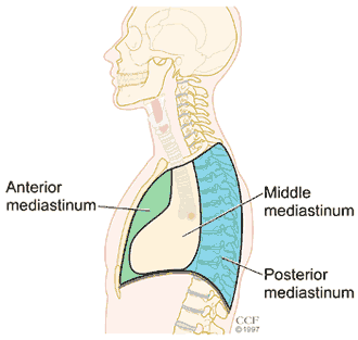
A mediastinal mass may be a solid growth, an enlarged lymph node, or a fluid-filled cyst. Some are found on a scan performed for an unrelated reason. Others cause cough, chest discomfort, shortness of breath, trouble swallowing, or hoarseness by pressing on nearby structures.
Not every mass is cancer
Cysts, enlargement of normal thymus tissue, and thyroid tissue extending into the chest are among the noncancerous possibilities. Some findings can be followed with imaging.
Location helps narrow the possibilities
The front of the mediastinum can contain thymic tumors, lymphoma, or germ cell tumors. Cysts and enlarged lymph nodes occur in other areas; nerve-related tumors often arise toward the back.
The scan alone does not always establish the diagnosis. The goal is to identify what the mass is before choosing observation, a biopsy, medication, or surgery.
Finding answers
How is a mediastinal mass evaluated?
- Review of symptoms and prior scans
- Your team asks about breathing, swallowing, weakness, fever, night sweats, and weight changes. Older images help show whether a finding is new or growing.
- CT scan
- A chest CT, usually with contrast when appropriate, shows the mass and its relationship to the heart, blood vessels, lungs, and other structures.
- MRI or PET scan in selected cases
- MRI can help distinguish a cyst from a solid growth or clarify nearby structures. PET may help assess certain tumors or their extent, but increased activity does not by itself prove cancer.
- Targeted blood tests
- Tests may look for clues to a germ cell tumor, thyroid-related condition, or myasthenia gravis. Not everyone needs every test.
Will I need a biopsy?
Sometimes. A needle biopsy or another sampling procedure may be important when lymphoma or another condition treated primarily with medication is suspected, or when treatment is planned before surgery.
A suspected thymoma that appears completely removable may be taken out without a separate biopsy first. In that setting, surgery can provide both the diagnosis and treatment while avoiding unnecessary disruption of the tumor capsule. Your team will explain why a biopsy is—or is not—the best next step.
Tell your team before a procedure
Report difficulty breathing when lying flat, new facial or neck swelling, or significant swallowing problems. A mass pressing on an airway or major blood vessel can affect the safety of sedation and anesthesia.
Thymic tumors
Thymoma and thymic carcinoma
A thymoma arises from cells in the thymus. Many grow slowly, but they can invade nearby tissues or return after treatment. A slow-growing thymoma still needs an appropriate treatment and follow-up plan. Thymoma can occur with myasthenia gravis, but having one does not necessarily mean you have the other.
Thymic carcinoma is a different, less common cancer of the thymus that generally behaves more aggressively. Expert review of the tissue helps distinguish these diagnoses.
How is treatment chosen?
For a tumor that can be completely removed, surgery is often the main treatment. The operation usually removes the tumor together with the thymus; nearby tissue or lymph nodes may also need to be removed depending on the findings.
If the tumor involves important surrounding structures, chemotherapy or other treatment may be recommended first. Radiation may be considered after surgery based on the tumor type, stage, and whether the edges of the removed tissue are free of tumor. Some patients need treatment without surgery.
At Lahey Clinic, the plan can involve thoracic surgery, medical oncology, radiation oncology, radiology, and pathology. The priority is complete and safe treatment—not simply the smallest incision.
Why follow-up matters
The final pathology report guides the next steps. Even after complete removal, thymoma can recur years later, so long-term imaging follow-up is important. Your team will give you a schedule based on your diagnosis.[1]
The thymus and the immune system
Myasthenia gravis: when can thymectomy help?
Myasthenia gravis (MG) is an autoimmune condition in which the immune system interferes with communication between nerves and muscles. It can cause fluctuating weakness, often worse with activity. Symptoms may include drooping eyelids, double vision, difficulty chewing or swallowing, changes in speech, or weakness of the arms, legs, and breathing muscles.
MG is not cancer. A neurologist helps confirm the diagnosis, often using antibody testing and nerve or muscle testing. Chest imaging checks for a thymoma.
MG with a thymoma
The tumor needs a treatment plan in its own right. When surgery is appropriate, removal of the thymoma and thymus is considered alongside careful management of the MG.
MG without a thymoma
Thymectomy may improve disease control and reduce medication needs in selected patients. It is not recommended for everyone with MG.
The strongest evidence for thymectomy without a tumor is in adults with generalized MG and acetylcholine receptor (AChR) antibodies, particularly younger adults. Age, duration and severity of illness, antibody results, response to medication, and surgical risk all matter. The role is less certain for weakness limited to the eyes and for some antibody-negative patients. It is generally not recommended for MuSK-antibody MG without a thymoma.[2]
Set realistic expectations
Thymectomy is not an immediate cure for MG. Improvement may develop over months to years, and medication may still be needed. Continue neurologic follow-up; do not reduce or stop treatment without your care team's guidance.
Surgical treatment
What does robotic thymectomy involve?
Dr. Servais uses robotic minimally invasive surgery for thymectomy and the removal of mediastinal masses when appropriate, including selected complex cases. He teaches and lectures on robotic thoracic surgical techniques.
During robotic thymectomy, Dr. Servais operates through small incisions using a magnified, three-dimensional view and precise instruments. He controls the instruments throughout the operation; the robotic system does not operate independently.
The thymus and the tissue needed for the planned operation are carefully separated from nearby structures. For MG, the goal includes removing surrounding fatty tissue that may contain additional thymus tissue. For a thymoma, keeping the tumor intact and achieving complete removal are important. The specimen is removed in a protective bag through an incision that may be enlarged as needed.
Can every operation be minimally invasive?
No. The tumor's relationship to the heart, major blood vessels, and other structures matters more than size alone. An open approach, such as an incision through the breastbone, may be the safest choice for some tumors. A planned robotic operation may need to be changed to an open operation if necessary for safety or complete removal.
For suitable patients, avoiding a breastbone incision can reduce the impact of the operation and support recovery. The appropriate approach is individualized. Alternatives may include observation, a biopsy, nonsurgical treatment, or a different surgical approach.
What risks should I understand?
Risks include bleeding, infection, pain, lung or anesthesia complications, and injury to nearby structures. The phrenic nerves, which control the diaphragm, run near the thymus; injury can affect breathing. Patients with MG also face a risk of worsening weakness or a need for breathing support after surgery. Your team will discuss the risks most relevant to you.
Learn more about Dr. Servais's robotic approach →Before your operation
Planning with your surgical and neurology teams
Preparation includes a review of imaging, medications, overall health, and heart or lung testing when needed. Tell the team about blood thinners, supplements, prior chest operations, and any weakness or difficulty swallowing.
If you have MG, the surgeon, neurologist, and anesthesia team coordinate your care. Symptoms should be well controlled before an elective thymectomy; surgery is generally postponed during a myasthenic crisis or poorly controlled disease. Selected patients receive intravenous immune globulin (IVIG) or plasma exchange before surgery.
Ask for a specific plan for your MG medicines, steroids, and other medications before and after the operation. Do not make these changes on your own. The team also plans postoperative monitoring and breathing support if needed.[2]
After surgery
Recovery and longer-term care
Hospital stay and recovery vary with the operation and your health. A temporary chest drain may be used. Walking, breathing exercises, and a tailored pain-control plan help you recover. Your discharge instructions should explain wound care, activity restrictions, driving, and when to return to work.
The postoperative visit includes a review of your pathology and any additional treatment or imaging needed. If you have MG, neurologic treatment continues even if recovery from the incisions is going well.[1]
What to know about chest tubes →Questions to bring to your visit
- What are the most likely explanations for my mass?
- Do I need more imaging or a biopsy before deciding on treatment?
- Is surgery intended to treat a tumor, help MG, or both?
- Can my operation be robotic, and what might require an open approach?
- How will my neurologist and surgical team coordinate my medications?
- What should I expect from recovery, pathology review, and long-term follow-up?
Sources and further reading
This guide provides general education for adults. Your care team will tailor recommendations to your diagnosis, symptoms, and health.
Patient resources, references & image credits
Patient resources
- National Cancer Institute: Thymoma and thymic carcinoma
- National Institute of Neurological Disorders and Stroke: Myasthenia gravis
Image credits
- Thymus anatomy illustration. Image provided by Elliot Servais, MD, for this guide.
Updated October 4, 2026.